The Life Span of the Red Blood Cell in Chronic Leukemia and…
The Life Span of the Red Blood Cell in Chronic Leukemia and Polycythemia Author(s):Nathaniel I. Berlin,John H.Lawrence and Helen C.Lee Source:Science,New Series, Vol. 114, No.2963 (Oct.12,1951), pp. 385-387 Published by: American Association for the Advancement of Science Stable URL: http://www.jstor.org/stable/1679527 Accessed:18-09-2016 03:01 UTC
JSTOR is a not-for-profit service that helps scholars, researchers, and students discover, use, and build upon a wide range of content in a trusted digital archive . We use information technology and tools to increase productivity and facilitate new forms of scholarship . For more information about JSTOR,please contact support@jstor.org.
Your use of the JSTOR archive indicates your acceptance of the Terms & Conditions of Use,available at http://about.jstor.org/terms
American Association for the Advancement of Science is collaborating with JSTOR to digitize, preserve and extend access to Science
No undesirable aftereffeets have been observed in animals given overdoses of the drug.
Several members of the class were tested for acute toxicity in rodents and dogs by oral and parenteral routes. Among these, 3-methyl-pentyne-ol-3 was notably low in toxicity. The acute oral LD for mice , rats, and guinea pigs,ranged from 600 to 900 mg/kg. The animals died in a state of coma. A few active compounds width low acute toxicity were tested for chronic toxicity in mice, rats,and dogs. 3-Methyl-pentyne-ol-3 at 200-300 mg/kg/day (approximately 70 times the recommended human dose)did not produce any gross or micropathological changes. Blood sugar levels, hemoglobin values, and erythrocyte, white blood cell, and differential counts were normal. Dogs that had reeeived chronically 3-methyl-pentyne-ol-3 per os showed normal renal function (phenolsulfonphthalein test).
In metabolic studies on 3-methyl-pentyne-ol-3 , the acetylenic hydrogen was used to identify and estimate the compound through the formation of the silver acetylide and the microdetermination of silver (o). Following the oral administration of 3-methyl-pentyne-ol-3(200 mg/kg) to adult dogs, 0.5-4.6% of the total dose was exereted in the urine during the first 24 hr; thereafter, only traces could be detected .In preliminary trials, no drug was detected in the urine obtained from three human subjects during a 60-hr period after a single oral dose of 100 mg. It was estimated that no more than 8% of the total dose could have been present in any 4-hr specimen. Following intravenous administration to dogs(200 mg/kg), it was found that the blood contained during the first 10 min 20% of the total dose; no drug could be detected in the blood at 2 hr . Analyses of rat tissues (brain,spleen,kidney,adipose,muscle,and liver) taken while the animals were still under the hypnotic action of 3-methyl-pentyne-ol-3 (800 mg/kg), revealed that adipose, muscle, and liver tissues together contained approximately 20% of the total dose. No unchanged compound was found in any of the organs or tissues when the effeets of the were no longer manifest. In ritro experiments in which 3-methyl-pentyne-ol-3 was added to whole blood from dogs and rats indicated that there was no breakdown of the compound; i.e., the acctylenie hydrogen was still present. llowever ,slices of kidney or liver, and to a lesser degree slices of brnin. Changed this moleeule in such a way as to render it nonreactive with the sliver rengent (9).
In a clinical study in 134 subjects, the majority of whom previonsly required barbiturates for sleep, 3-methyl pentyne-ol-3 was found to be highly active, without toxie effect,and free from undesirable side actions (10). The effective oral dose in adults was 200-300 mg. Sleep was brought about in the majority of patients in less than 1/2 hr. The patients who received 3-methyl-pentyne-ol-3 experientced restful sleep and had no “hangover” upon awakening. A number of patients have been given doses of the compound for more than 6 months without any untoward effects Complete blood counts, urinalyses, blood sugar, blood urea nitrogen, cratinine, total serum protein, albumin, globulin, phosphorous, alkaline phosphotase, total cholesterol, free and combined cholesterol, and in addition, the icterus index, Van den Bergh, thymol turbidity, or cephalin flocculation values were determined, before and after medication with 3-methyl-pentyne-ol-3. These clinical laboratory tests indicated that there were no pathologieal changes attributable to the drug.
The Life Span of the Red Blood Cell in Chronic Leukemia and Polycythemia 1,2
Nathaniel I. Berlin, John H. Lawrence and Helen C. Lee
Donner Laboratory of medical pbysics, University of califormia, Berkeley
The anemia associated with leukemia and meoplastie diseases is still not completely understood; infiltration of the erythropoietic tissue, has been postulated as a signifieant factor in the pathogenesis of the anemia. IIowever, Huff and co-workers (1) have shown with radionetive iron that in some patients with leukemia the rate of production of red blood cells was greater than the 0.8% per day that would be anticipated if the cells lived a normal life span of approximately 120 days (2). These same workers showed that in polycythemia vera the rate of production of red cells was much greater than 0.8% per day. These findings may be explained by postulating a decrease in thelife span of the red blood cells in these diseases. Using N15-labeled glycine, which Shemin and Ritten-berg(2) demonstrated to be a specific precursor of the porphyrin of hemoglobin, London et al. (3) found a normal life span of the red blood cell in a single case of polycythemia vera.
Since C14-labeled glycine has been shown to be satis-factory for the labeling of rat hemoglobin (4,5) and for the determination of the life span of the red blood This study was supported in part by the Atomte Energy Commlssion and the U.S Puble Health Servler .Presentel before the American Feleration for Clinical Research,Atlantic City, N,J.,May 1,1951.
Cell of the dog (5,6) and rat (7) ,studies (8) utilizing C14-methyl-labeled glycine were undertaken to deter-mine the red blood cell life span in patients with leukemia and polyeythemia.
Three patients with chronic leukemia,one with lymphatic and two with myelogenous, and two patients with polyeythemia vera were given intravenously 8.6mg of glycine 2-C14 containing 100μc of C14 .Blood samples were drawn at frequent intervals, and the hemoglobin was extracted by a modification of the method of Drabkin (9). The plasma was removed by centrifugation, the cells were washed three times with saline and then lysed with distilled water; the lipids were extracted with toluene . After standing overnight, the solution was centrifuged and an aliquot of the clear hemoglobin solution was dried in vacuo and combusted to CO2 by the method of Van Slyke and Folch (10). This was precipitated was BaCO2; a l-g aliquot was converted to CO2 and the specific radioactivity measured in a 100-ml ionization chamber (11), using a vibrating reed electrometer and recording poten-tiometer.
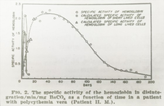
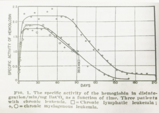
Figs. 1 and 2 show the graph for the specific activity of hemoglobin as a function of time in the three patients with leukemia (Fig.1) and one patient with polyeythemia vera (Fig.2).

Fig.1 shows that the red blood cells in one patient with chronic lymphatic leukemia in clinical and hema-tologieal remission at the time of these studics had a normal mean life span (100 days),and that in the tow patients with myelogenous leukemia who were anemic there is considerable shortening of the mean life span of the red blood cell, from a normal of approximately 120 ,to 71 and 76 days,respectively . If the bone marrow in these patients continued to produce a normal number of red blood cells per days, it is then evident that these paticnts will become anemic. Therefore, in leukemin, a significant factor in the pathogenesis of the ancmia is the decrease in the life span of the red blood cell.

Fig. 2,which shows the specifie activity of the hemoglobin as a function of both patients with polyeythemia vera, may be analyzed in the following manner: The graph from a period of 40-200 days is qualitatively similar to that of the normal and to the leukemic, so that in these individuals there is at least one class of red blood cells that had an almost normal life span; the rapid initial rise and fall in the first 15 days may be analyzed by considering this as due to the delivery into the peripheral eireulation of a second class of red blood cells with a life span of only a few days. If the long-lived cells are delivered into the peripheral eireulation at a rate consistent with a first-order process, which is what Shemin and Ritten-berg (2) found, then , by determining the rate graph-ically in the interval 20-40 days after administration of the radioactive glycine, the curve for the specific activity of this class of cell may be extrapolated to the origin. The difference. Between the extrapolated curve for the long-lived cells and the observed specific activity is due to the presence of the short-lived cells, and so the curve for the short-lived cells may be constructed. It is assumed in the above diseussion that in both elasses of red blood cells that the removal of the hemoglobin and the red blood cell from the circu-lation is the same process.
Huff and his co-workers (1),in their radioactive iron studies, have prescnted evidence which “may indicate that the longevity of red cells in polycythemia vera is significantly shortened.” The present results show that such short-lived cells do exist, but that there is also present a class of cells with a normal life span. It is the rapid turnover of these short-lived red blood cells that is largely responsible for the greatly increased rate of utilization of iron for the formation fo hemoglobin in this disease.
Further studies involving particularly the separation of the hemoglobin into the constituent porphyrin and globin moictics are in progress.
These studies may necessitnte a revision of our present concept of the mechanism of the anemia occurring in neoplastic disease.
Tissue Cytochrome c and Prevention of Experimental Atherosclerosis
J.Mardones,J. Monsalve,M. Vial,
And M. Plaza de los Reyes
It has been shown that the protective action of iodides on the experimental atherosclerosis induced in rabbits by cholesterol-rich dicts is exerted through the thyroid, since iodides are not effective in the absence of the gland (1,2) or when given simultaneously with thiourea (2).
Drabkin (3) has pointed out that there is a clear positive correlation between the thyroid activity and the content of cytochrome c in the tissues. Consequently it seemed important to investigate whether changes will occur in the cytochrome c content of tissues of rabbits on a cholesterol-rich diet with potassium iodide, in the presence and in the abscnce of thyroid gland. Rabbits on a cholesterol-rich diet (nearly 0.5 g cholesterol, in the form of cattle spinal cord. Daily) and with constriction of the upper abdominal aorta inducing hypertension, which acts synergistically with diet in producing atherosclerosis (4), were divided into 4 groups:one control, another with thyroidec-tomy, and two on protective doses of potassium iodide (0.3 g orally every other day), one of them thyroidectomized. The animals were sacrificed 120 days after starting the diet. The development of atheroselerosis was judged macroscopically and evaluated on a scale of 0 to 10 (4). The eytochrome c of liver and kidney was extracted by the method of Potter and Du Bois (5) and determined spectrophotometrically according to Rosenthal and Drabkin (6).
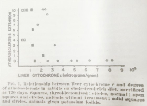
The results on liver eytochrome c are given in Fig.1. The changes in kidney eytochrome c were similar to those in the liver.
The correlation coefficient between liver eytochrome c and development of atherosclerosis is r = -0.533, with t = 2.88, a value regarded as statistically significant.

These findings confirm, in the rabbit, Drabkin’s results in the rat (3) of the influence of the thyroid gland and thyroxine upon the leven and content of eytochrome c in tissues. It is deduced from the data that the action of iodide may be one of stimulation of the thyroid gland, since the concentration of cellular eytochrome c is increased when the drug is administered to animals with thyroid. The results furthermore suggest that the augmentation of cellular eytochrome c must be considered as a factor in the prevention of the experimental atherosclerosis by means of potassium iodide.


